
3D ultrasounds have become an increasingly common part of prenatal care and provide expectant parents with a more detailed view of their developing child. This technology goes beyond the scope of traditional 2D ultrasounds. They can offer a deeper level of visualization for both parents and OBGYNs. But what exactly is a 3D ultrasound, how does it differ from traditional scans, and what happens during the process?
What Is a 3D Ultrasound?
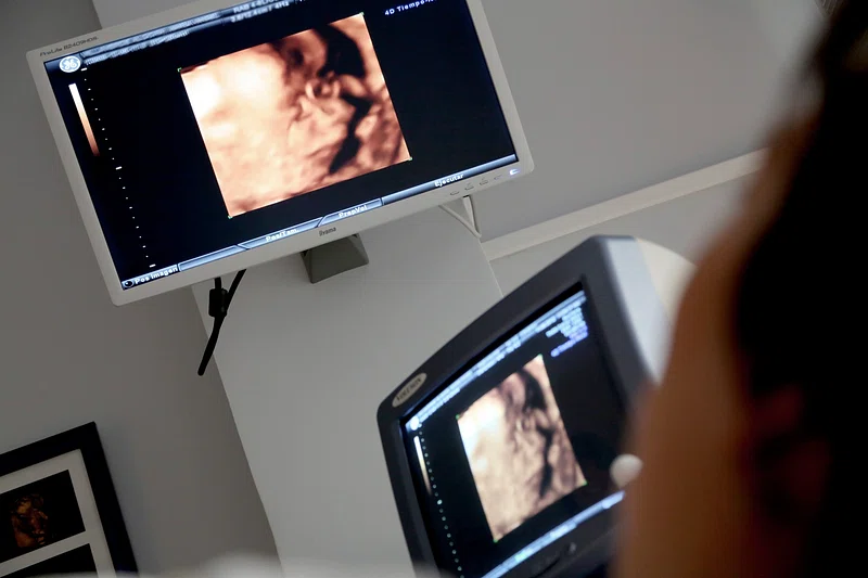
A 3D ultrasound is a medical imaging technique that uses sound waves to create three-dimensional images. It is typically performed by an OBGYN or another medical professional. Traditional 2D ultrasounds provide flat, grayscale images. 3D ultrasounds combine multiple angles from the sound waves to form detailed, tangible visuals of the fetus. These images offer depth and clarity, presenting a lifelike picture of the baby. While 3D ultrasounds are not a standard part of routine prenatal care, they are often used in cases where deeper analysis may be beneficial. They might offer enhanced visualization for observing conditions or confirming findings from earlier ultrasounds.
Are They More Useful Than Traditional Ultrasounds?
While both 2D and 3D ultrasounds serve their own purposes, each has unique advantages. Traditional 2D ultrasounds remain the standard diagnostic tool for prenatal care. 3D ultrasounds, on the other hand, add a layer of visualization that brings more depth to the assessment. This type of imaging allows healthcare providers to examine certain structural details of the fetus more accurately. Conditions related to the face may be easier to detect because of this technology’s advanced depiction capabilities. The detailed images can offer a more intimate connection, showcasing facial features that a standard 2D scan would not effectively show.
What Does the Process Involve?
The procedure is noninvasive and will usually begin with the application of a specialized gel to the abdominal area. This gel serves as a medium for sound wave conduction. A probe is then applied to the abdomen. The probe emits sound waves that travel into the body and reflect back to create an image. For 3D ultrasounds, the equipment captures multiple angles and generates a three-dimensional image.
Once the procedure is completed, parents may receive printed images or digital downloads of the 3D scans. It is worth noting that while 3D ultrasounds can serve as a keepsake for parents, their primary aim remains medical. The images serve as a diagnostic supplement to assess the baby’s development and provide meaningful insights when necessary.
Seek Out an OBGYN That Can Perform This Test
A 3D ultrasound offers unique benefits for both expecting parents and healthcare providers. Its ability to produce detailed, lifelike images and clearly showcase certain developmental details makes it an effective complement to traditional 2D ultrasounds. If you wish to learn more about 3D ultrasounds or would like to schedule one, consult an OBGYN who provides this service. They can make sure that this procedure aligns with your overall prenatal care plan while providing you with an informative and meaningful experience. By selecting a trusted healthcare provider, you can gain enhanced insight into your baby’s development.
- Choosing the Right Plastic Surgeon for Your Cosmetic Procedure
- Understanding Different Types of Laser Treatments for Skin Rejuvenation
- Why a Family Dentist is Key for Maintaining Oral Health
- The Benefits of Regular Visits to a Wellness Spa
- Exploring the Emotional and Psychological Triggers of Eating Disorders
Leave a Reply