
X-rays play a significant role in modern medical diagnostics, particularly in identifying and understanding bone-related conditions. This diagnostic tool is a quick and effective way to visualize what cannot be seen externally. Here is more information on the function of X-rays and how they contribute to the diagnosis of bone conditions:
What Is an X-ray?
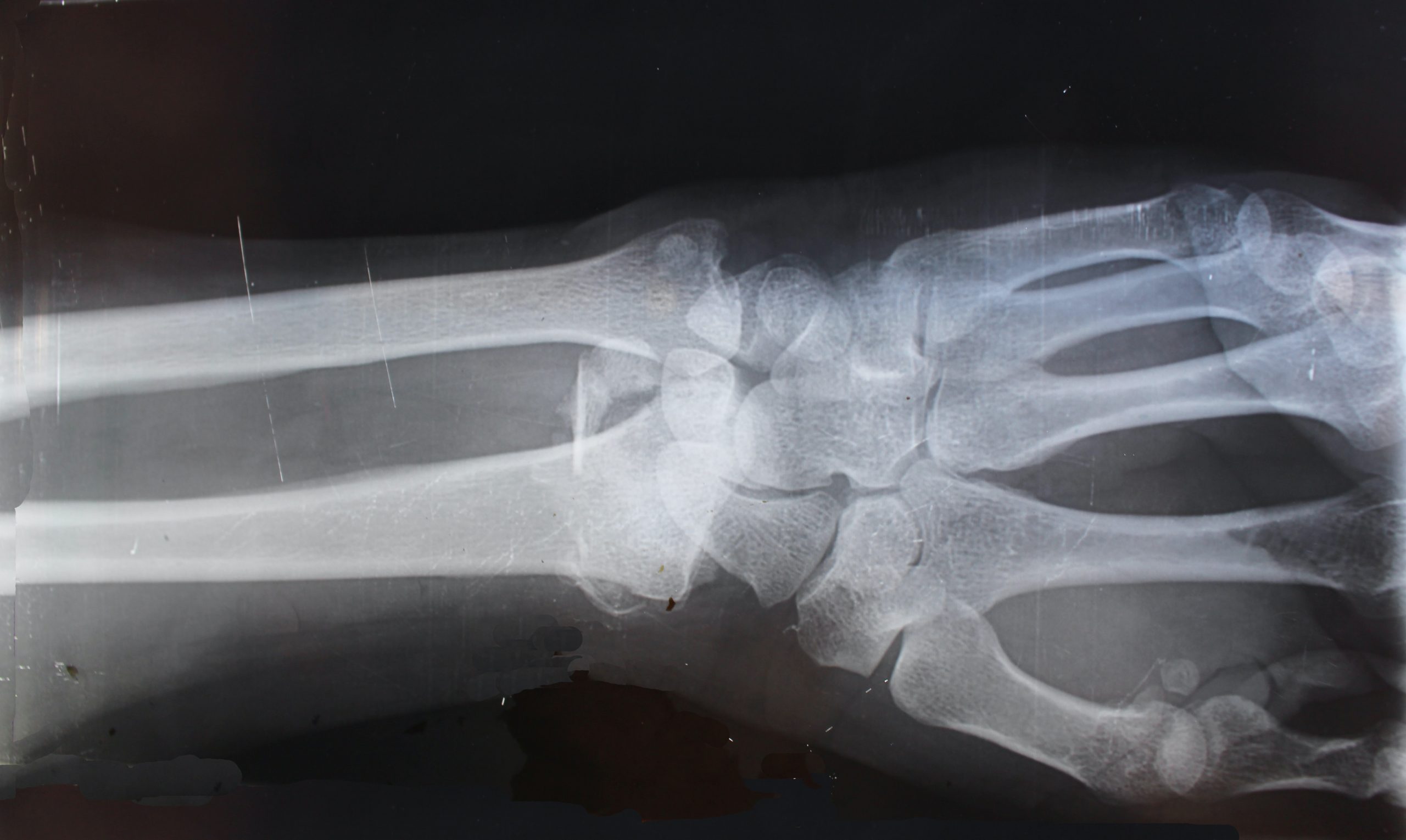
An X-ray is a diagnostic imaging technique that creates visual representations of the inside of the body. It is used to capture images of bones, joints, and certain tissues. X-rays rely on electromagnetic waves, which pass through the body and produce images based on variations in density. These images allow healthcare providers to detect abnormalities or underlying issues within the skeletal system.
X-ray images appear in grayscale. Bones and other dense materials, which block X-ray waves, appear white. Softer tissues that absorb less radiation, such as muscles and fat, appear in shades of gray. This contrast enables the discernment of structural details and abnormalities.
How Does It Work?
X-rays work by emitting electromagnetic radiation through a targeted area of the body. A machine directs controlled doses of X-ray beams toward the area being examined. These beams pass through the body and are absorbed at different rates depending on tissue density.
A detector on the opposite side captures the remaining radiation and converts it into an image. Denser materials, such as bones, absorb more radiation, allowing physicians to visualize their structure, alignment, and condition. The procedure itself is painless and typically takes only minutes to complete.
What Are Bone Conditions?
Bone conditions refer to a variety of disorders that affect the skeletal system. These conditions can disrupt normal bone structure, function, or density. Examples include fractures, osteoporosis, arthritis, infections, and deformities.
Fractures are breaks in a bone caused by trauma, overuse, or conditions that weaken bone strength. Osteoporosis involves a decrease in bone density, increasing the risk of fractures. Arthritis affects joint function and causes inflammation, while infections can lead to more complex bone deformities. Bone conditions can vary widely in severity, ranging from minor issues that only require observation to significant disorders that need medical intervention.
How Are They Diagnosed?
X-rays are a primary diagnostic tool used to identify a range of bone conditions. Physicians may recommend X-rays as a first-line procedure due to their ability to provide detailed images of skeletal structures. During an X-ray, the affected area is exposed to radiation, and images are captured to assess the condition of underlying bones and joints.
Fractures can be diagnosed by examining the alignment and continuity of bones in X-ray images. Misaligned or broken sections of bone are clearly visible, helping physicians determine the severity of the injury. For conditions like osteoporosis, X-rays reveal thinning or porous bones, while arthritis may show joint space narrowing or the development of bone spurs.
Seek a Diagnosis Now
Diagnosing bone conditions early is a valuable step toward effective treatment and improved quality of life. X-rays provide a reliable, fast, and accessible method for detecting bone-related issues. Whether you’re dealing with persistent pain, limited mobility, or recent trauma, consulting an orthopedic specialist for an X-ray can clarify the situation and guide the next steps. If you’re facing symptoms of a bone condition, schedule an appointment for an evaluation now.
- Choosing the Right Plastic Surgeon for Your Cosmetic Procedure
- Understanding Different Types of Laser Treatments for Skin Rejuvenation
- Why a Family Dentist is Key for Maintaining Oral Health
- The Benefits of Regular Visits to a Wellness Spa
- Exploring the Emotional and Psychological Triggers of Eating Disorders
Leave a Reply